Skeletal Muscle Fibrosis and Aging: When Support Tissue Gets in the Way
Muscle needs connective tissue, but excess fibrosis can limit repair and movement. Explore how aging shifts the extracellular matrix and what supports muscle quality.
Table of Contents
DISCLAIMER
This article is for informational purposes only and does not constitute medical advice. The statements in this article have not been evaluated by the FDA. The information presented is based on published research and should not be used as a substitute for professional medical guidance. Consult your physician before starting any supplement or health protocol.
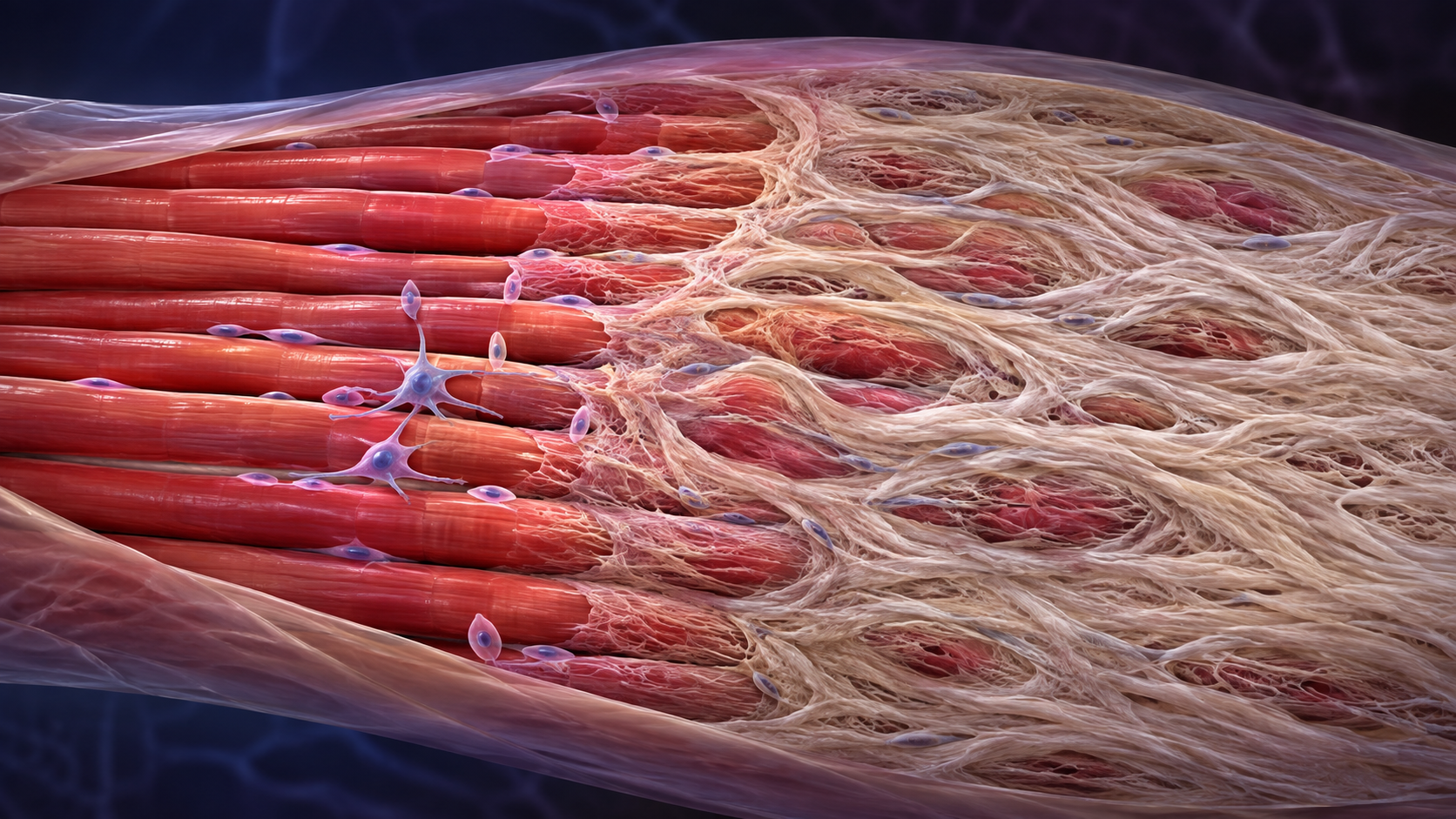
Muscle is not made of contractile fibers alone. Every fiber sits inside an extracellular matrix that organizes tissue, transmits force, carries vessels and nerves, and helps stem cells interpret injury.
With aging, inactivity, repeated injury, and some diseases, this support network can become excessively dense or poorly remodeled. That process—fibrosis—may reduce muscle quality even when a scan shows a reasonable amount of muscle mass.
The matrix is part of the machine
Collagens, laminins, proteoglycans, and other matrix components form layers around fibers, bundles, and the entire muscle. They provide a scaffold strong enough to transmit force while remaining compliant enough for contraction and stretch.
After injury, temporary matrix deposition stabilizes the area. Immune cells, fibro-adipogenic progenitors, muscle stem cells, and local signals coordinate cleanup and rebuilding. In a successful repair, much of the provisional material is reorganized as new fibers mature.
Fibrosis occurs when deposition outpaces removal or when repair signaling stays active. More matrix is not automatically stronger tissue. Excess collagen can separate fibers, change stiffness, interfere with cell movement, and alter how force spreads across the muscle.
Why aging can tilt repair toward scar
Aging changes inflammatory signaling, blood supply, stem-cell behavior, and the biochemical environment of the matrix. Older muscle may resolve inflammation less efficiently after injury. Fibro-adipogenic progenitors that normally support repair can contribute to excess connective tissue or fat when regulatory signals are disrupted.
Advanced glycation end products can form cross-links in long-lived matrix proteins, increasing stiffness. Physical inactivity removes the repeated mechanical signals that help maintain tissue organization. Neuromuscular loss and reduced loading can further change which regions are actively maintained.
These processes overlap with sarcopenia but are not identical to it. Sarcopenia is defined clinically through low strength, reduced muscle quantity or quality, and impaired performance. Fibrosis is one possible contributor to poor quality, alongside fat infiltration, mitochondrial changes, motor-unit loss, and altered architecture.
Why fibrosis is difficult to measure
A biopsy can quantify collagen and reveal tissue organization, but it samples a tiny region and is invasive. Magnetic resonance methods, ultrasound texture, and elastography may provide indirect information about composition or stiffness, yet stiffness is influenced by contraction, hydration, fat, and measurement position.
Blood markers of collagen turnover reflect activity across multiple tissues, not one muscle. A consumer body-composition scan cannot isolate microscopic scar tissue. This is why fibrosis remains more useful as a biological explanation than as a routine longevity metric.
Persistent weakness, pain, swelling, or loss of function should be evaluated for common neurological, orthopedic, inflammatory, and metabolic causes. Labeling the problem “fibrosis” without an assessment can delay useful care.
Training muscle and its support tissue
Progressive resistance training remains the most direct way to preserve strength and challenge muscle tissue. Aerobic activity supports circulation and metabolic health, while adequate protein and total energy help the body adapt to training.
Loading should rise gradually, especially after injury or prolonged inactivity. Recovery time matters because repeated damage without adequate repair can be counterproductive. Conditions such as diabetes, vascular disease, and chronic inflammatory illness deserve appropriate treatment rather than an unproven anti-fibrosis supplement.
Healthy muscle is a collaboration between cells and scaffold. The goal is not to eliminate connective tissue, but to maintain a matrix that is strong, adaptable, and capable of making room for repair.
Frequently Asked Questions
Is all connective tissue in muscle harmful?
Can a routine scan diagnose muscle fibrosis?
Can exercise reverse muscle fibrosis?
Sources
Stay Updated on Longevity Science
Weekly research digests. No spam, unsubscribe anytime.
Related Articles

Ankle Power and Healthy Aging: The Push-Off That Keeps Walking Efficient
Ankle plantar-flexor power supports walking speed and balance recovery. Learn why push-off changes with age and how strength can be trained safely.
9 min read
Arterial Calcification and Vascular Aging: Reading the Risk Signals
Arterial calcification can reveal accumulated vascular risk. Learn what calcium scores mean, why calcification develops, and where testing has limits.
10 min read
Arterial Stiffness and Vascular Age: A Longevity Evidence Guide
Understand arterial stiffness vascular age longevity links, key studies, measurement limits, and evidence-based lifestyle implications for healthy aging.
11 min read