Perivascular Fat and Aging: The Tissue Talking to Your Arteries
Perivascular adipose tissue can help or hinder vessel function. Learn how this active fat depot changes with aging, inflammation, and metabolic health.
Table of Contents
DISCLAIMER
This article is for informational purposes only and does not constitute medical advice. The statements in this article have not been evaluated by the FDA. The information presented is based on published research and should not be used as a substitute for professional medical guidance. Consult your physician before starting any supplement or health protocol.
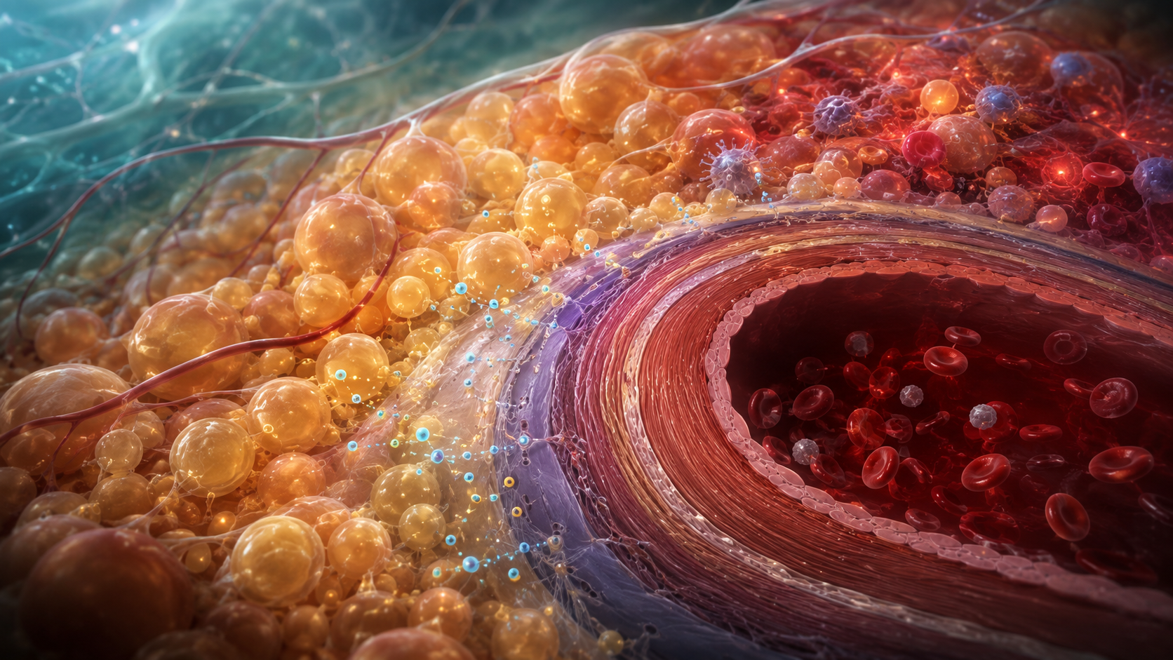
Arteries do not sit alone. Many are wrapped in perivascular adipose tissue, a local fat depot once treated as passive packing material. Researchers now view it as a signaling organ that communicates with the vessel wall next door.
In a healthy state, this tissue can release factors that support relaxation and buffer mechanical stress. With metabolic disease and aging, its signals may shift toward inflammation, oxidative stress, and impaired vascular function.
A fat depot with a local audience
Most endocrine signals travel through the bloodstream. Perivascular fat can also signal directly across short distances to smooth-muscle and endothelial cells. This local route is often described as paracrine communication.
The tissue varies by location. Fat surrounding the aorta is not identical to fat near coronary or small resistance vessels. Cell type, developmental origin, temperature response, and metabolic activity can differ, so findings from one vascular bed cannot automatically be applied to another.
Healthy perivascular tissue may release adiponectin, nitric-oxide-related signals, and other mediators that reduce contraction. It also provides physical support. The important distinction is therefore not simply how much fat exists, but what state the tissue is in.
How aging and metabolic stress alter the conversation
With obesity, insulin resistance, smoking, and some forms of hypertension, fat cells may enlarge while immune-cell activity and inflammatory signaling increase. Oxidative stress can reduce nitric oxide availability, making it harder for vessels to dilate normally.
Aging can add cellular senescence, mitochondrial dysfunction, fibrosis, and altered immune regulation. In some depots, characteristics associated with energy-burning brown or beige fat may decline. The resulting tissue may become less metabolically flexible and more inflammatory.
Signals move in both directions. A stressed vessel can influence nearby fat, while dysfunctional fat can affect the vessel wall. This feedback may contribute to endothelial dysfunction and atherosclerotic biology, but it does not mean that perivascular fat alone causes cardiovascular disease.
What imaging can reveal—and what it cannot
Computed tomography and magnetic resonance imaging can estimate fat volume in selected regions. Researchers also study tissue density or inflammation-related imaging features around coronary arteries. These methods are valuable for research and, in defined settings, risk assessment.
They do not provide a universal “artery fat age.” Scanner protocols, anatomy, disease status, and the vascular bed all affect interpretation. Imaging also cannot fully describe the molecules released by living tissue without additional biological measurements.
Routine cardiovascular decisions still rely on established information such as blood pressure, cholesterol, diabetes status, smoking, symptoms, family history, and clinically indicated imaging. A specialized perivascular-fat metric should not displace those basics.
Improving the metabolic neighborhood
Regular physical activity, avoidance of smoking, adequate sleep, and management of blood pressure, lipids, and glucose support vascular and adipose health together. For people with excess body fat, sustainable weight reduction can lower systemic inflammatory and metabolic stress, though no home strategy selectively targets fat around one artery.
Medications that reduce cardiovascular risk should be chosen for established indications with a clinician. Supplements marketed as vascular-fat burners have not been shown to rejuvenate perivascular tissue throughout the body.
Perivascular adipose tissue changes the mental picture of an artery. Vascular aging is not confined to the inner lining or the plaque we can image. It also involves the surrounding neighborhood—and the quality of the biological conversation happening across the vessel wall.
Frequently Asked Questions
Is perivascular fat the same as plaque?
Can a waist measurement show perivascular fat?
Should all fat around arteries be removed?
Sources
Stay Updated on Longevity Science
Weekly research digests. No spam, unsubscribe anytime.
Related Articles

Ankle Power and Healthy Aging: The Push-Off That Keeps Walking Efficient
Ankle plantar-flexor power supports walking speed and balance recovery. Learn why push-off changes with age and how strength can be trained safely.
9 min read
Arterial Calcification and Vascular Aging: Reading the Risk Signals
Arterial calcification can reveal accumulated vascular risk. Learn what calcium scores mean, why calcification develops, and where testing has limits.
10 min read
Arterial Stiffness and Vascular Age: A Longevity Evidence Guide
Understand arterial stiffness vascular age longevity links, key studies, measurement limits, and evidence-based lifestyle implications for healthy aging.
11 min read