The Osteocyte Network and Bone Aging: How Buried Cells Sense Every Step
Osteocytes form a communication network inside bone that senses load and coordinates remodeling. Explore how this system changes with age and disuse.
Table of Contents
DISCLAIMER
This article is for informational purposes only and does not constitute medical advice. The statements in this article have not been evaluated by the FDA. The information presented is based on published research and should not be used as a substitute for professional medical guidance. Consult your physician before starting any supplement or health protocol.
Bone looks inert because most of its cells are hidden inside mineral. Yet those cells form a branching sensory network that experiences bending, pressure, and fluid movement whenever a person walks, lifts, or lands.
These embedded cells are osteocytes. They help translate mechanical use into instructions for bone formation and resorption. Changes in their number, connections, and signaling may contribute to the declining ability of older bone to repair damage and adapt to load.
A Living Network Inside Mineral
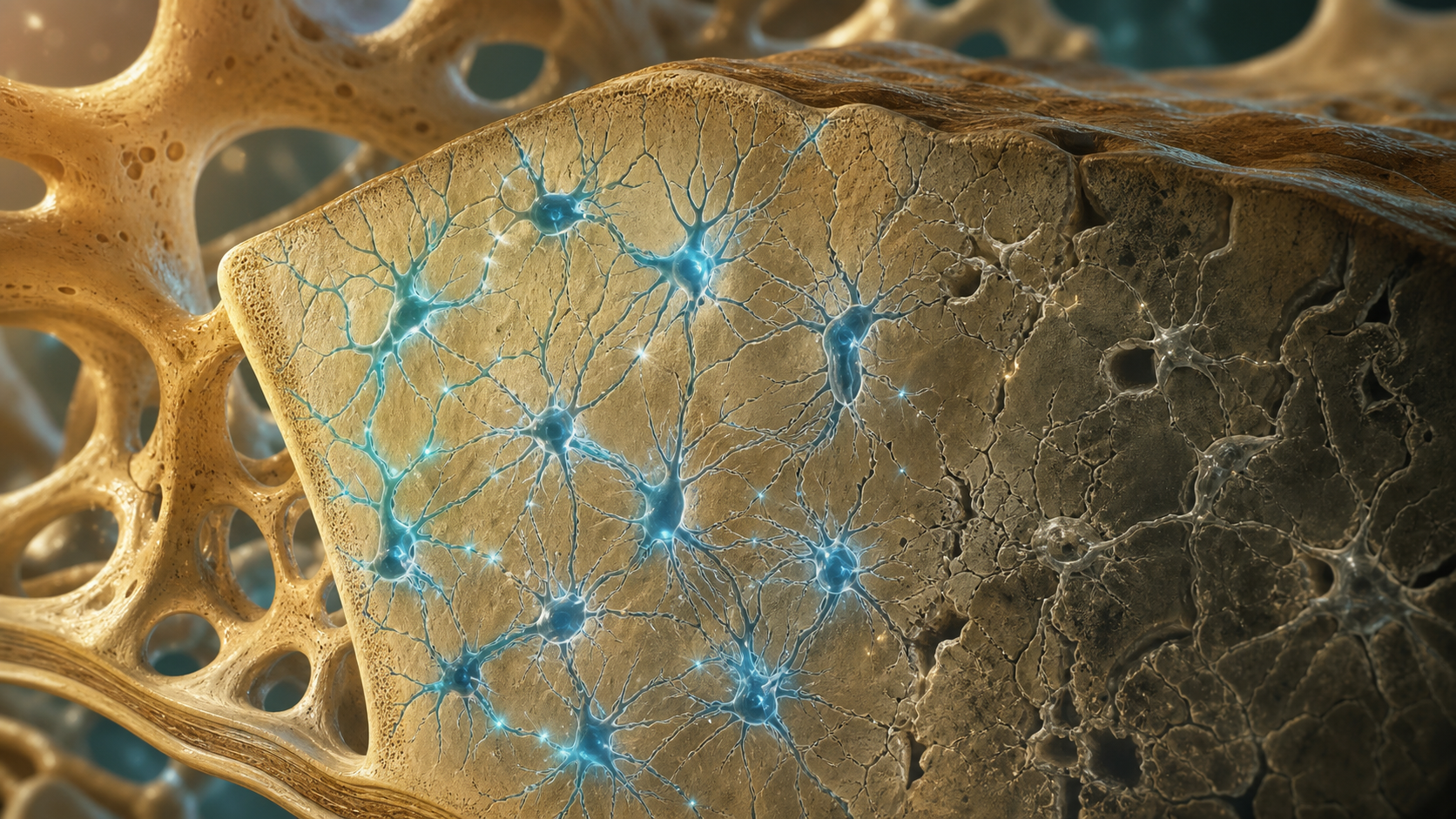
An osteocyte occupies a tiny cavity called a lacuna. Slender cell processes extend through canaliculi and connect with neighboring osteocytes and cells on bone surfaces. The resulting lacunar-canalicular network carries signals across tissue that would otherwise seem sealed in stone.
Mechanical loading deforms bone by a very small amount. That deformation moves fluid through the microscopic channels, creating forces the cells can detect. Osteocytes respond through calcium signals, electrical and chemical communication, and changes in regulatory proteins.
One important signal is sclerostin, which generally restrains bone-forming pathways. Mechanical loading can reduce sclerostin signaling in relevant contexts, while unloading can shift the balance toward bone loss. Osteocytes also influence RANKL and other signals that regulate bone-resorbing osteoclasts.
What Changes With Age
With age, some osteocytes die and empty lacunae accumulate. The network may become less connected, while the surrounding matrix changes in mineralization, collagen cross-linking, and microdamage. Surviving cells can also adopt altered inflammatory or senescence-associated signaling.
The consequences are not limited to bone density. Bone quality depends on architecture, material properties, turnover, damage repair, and geometry. Two people with similar density measurements can have different fracture risks because these features differ.
Hormonal change, reduced activity, kidney disease, glucocorticoid exposure, nutrition, and other conditions can affect the same system. Osteocyte aging is therefore one contributor within a larger remodeling problem, not a single master cause of osteoporosis.
Loading Is Information, Not Just Force
Bone responds most strongly to loading that is meaningfully different from its routine environment. Weight-bearing activity, resistance exercise, and—when safe—brief higher-rate impacts can provide osteogenic signals. Muscle contractions matter because they pull on bone even without jumping.
More force is not always better. Bone needs recovery, and a structure weakened by osteoporosis or an existing lesion may fail before it adapts. Exercise selection should account for fracture history, balance, pain, joint replacement, and medical treatment.
Disuse sends the opposite message. Bed rest, immobilization, and prolonged microgravity reduce normal mechanical input and can accelerate bone loss. Even then, the response varies by skeletal site: a forearm, spine, and hip do not share identical loading histories.
What Can Be Measured and Treated
Standard DXA scanning estimates areal bone mineral density. It does not count living osteocytes or map their microscopic connections. High-resolution imaging and bone biopsy can reveal more structure in research or selected clinical settings, but no routine consumer test reports “osteocyte age.”
Clinically, fracture prevention combines risk assessment, adequate nutrition, suitable physical activity, fall reduction, and medication when indicated. Antiresorptive and bone-forming therapies act on remodeling pathways in different ways; treatment decisions require individual medical evaluation.
The osteocyte network changes how we think about a skeleton. Bone is not simply a mineral bank that becomes emptier with time. It is a sensory organ that continually interprets use, damage, hormones, and metabolism—and its long-term health depends on keeping that conversation active and safe.
Frequently Asked Questions
What is an osteocyte?
Do osteocytes build bone directly?
Can impact exercise improve every aging bone?
Sources
Stay Updated on Longevity Science
Weekly research digests. No spam, unsubscribe anytime.
Related Articles

Ankle Power and Healthy Aging: The Push-Off That Keeps Walking Efficient
Ankle plantar-flexor power supports walking speed and balance recovery. Learn why push-off changes with age and how strength can be trained safely.
9 min read
Arterial Calcification and Vascular Aging: Reading the Risk Signals
Arterial calcification can reveal accumulated vascular risk. Learn what calcium scores mean, why calcification develops, and where testing has limits.
10 min read
Arterial Stiffness and Vascular Age: A Longevity Evidence Guide
Understand arterial stiffness vascular age longevity links, key studies, measurement limits, and evidence-based lifestyle implications for healthy aging.
11 min read