Myosteatosis and Muscle Aging: Why Fat Inside Muscle Matters
Myosteatosis and muscle aging are linked to weaker function and metabolic risk, but imaging, causes and practical interpretation require context.
Table of Contents
DISCLAIMER
This article is for informational purposes only and does not constitute medical advice. The statements in this article have not been evaluated by the FDA. The information presented is based on published research and should not be used as a substitute for professional medical guidance. Consult your physician before starting any supplement or health protocol.
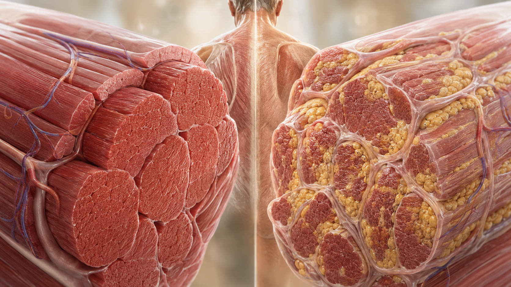
Two people can have similar amounts of muscle but very different muscle quality. Myosteatosis and muscle aging describe part of that difference: fat accumulates between muscle groups, between fibers and sometimes inside muscle cells, where it may interfere with force production and metabolism.
Myosteatosis is not visible marbling in the everyday sense, nor is it simply obesity. It is an imaging and biological pattern that researchers increasingly connect with frailty, insulin resistance, surgical risk and declining mobility.
What myosteatosis means
Skeletal muscle stores small lipid droplets as a normal energy reserve. Endurance-trained athletes can carry substantial intramuscular lipid while remaining highly insulin sensitive—sometimes called the athlete’s paradox. The concern arises when fat infiltration appears alongside poor mitochondrial capacity, inflammation, weakness or reduced physical activity.
Researchers distinguish intermuscular adipose tissue, which lies between muscle groups, from intramuscular fat within fascial compartments and lipid inside muscle fibers. Studies do not always use these terms consistently, which complicates comparisons.
Why it tends to increase with age
Several processes may converge. Physical activity often falls, muscle fibers shrink, insulin signaling changes and stem-cell-mediated repair becomes less efficient. Hormonal shifts and chronic low-grade inflammation can favor fat storage. After injury or prolonged bed rest, connective tissue and fat may replace some contractile tissue.
The relationship runs both ways. Reduced movement can promote fat infiltration, while lower-quality muscle can make movement harder. Chronic disease, corticosteroid exposure and inadequate protein intake may add to the cycle.
Muscle quantity is not muscle quality
DXA scans estimate lean mass but cannot fully show what that tissue contains. A person may retain acceptable lean mass while strength or power declines. Grip strength, chair-rise performance and walking speed therefore provide functional information that a body-composition number cannot.
This helps explain why modern sarcopenia definitions increasingly combine muscle quantity with strength and performance rather than relying on mass alone.
How researchers measure it
CT scans estimate tissue density in Hounsfield units. Fat lowers muscle attenuation, so lower-density muscle can indicate greater lipid infiltration. MRI can separate fat and water more directly, while proton spectroscopy can examine lipid inside cells. Ultrasound echo intensity is cheaper and portable but depends heavily on technique and device settings.
No single threshold works everywhere. Cutoffs vary by sex, ethnicity, muscle group, scanner settings and study population. An incidental CT comment about fatty muscle should be interpreted with the broader clinical picture rather than treated as a standalone diagnosis.
What the research suggests
A National Institute on Aging workshop report led by Correa-de-Araujo in 2020 described myosteatosis as an important component of skeletal muscle function deficit and emphasized the need for standardized definitions. The report connected imaging signals with metabolic and mobility outcomes while warning that methods remain heterogeneous.
A 2023 review by Axelrod and colleagues placed muscle fat within sarcopenic obesity, where low muscle function and excess adiposity interact. A 2024 imaging review by Garcia-Diez and colleagues summarized CT, MRI and ultrasound approaches and their diagnostic limits.
Observational studies often associate lower muscle density with poorer strength, falls, complications after major surgery and mortality. Those associations do not prove that removing muscle fat alone would reverse risk. Myosteatosis may partly be a marker of inactivity or systemic illness.
What may support better muscle quality
Progressive resistance training is the most direct way to challenge contractile tissue. Aerobic exercise can improve mitochondrial function and insulin sensitivity. Programs combining the two may address both strength and metabolic health.
Adequate dietary protein, sufficient total energy and correction of vitamin deficiencies may matter, particularly for older adults at risk of malnutrition. Weight loss requires care: aggressive calorie restriction can reduce both fat and muscle. Clinician or dietitian guidance is useful when frailty, kidney disease or recent illness is present.
Sleep, smoking cessation and management of diabetes or cardiovascular disease support the same system. There is no supplement proven to selectively remove fat from aging muscle.
Limitations and future research
Researchers still need harmonized cutoffs and longitudinal trials linking changes in imaging to changes in function. Muscle fat may also behave differently across the thigh, calf and trunk. Future work should separate harmless energy storage from lipid species associated with cellular stress.
Better opportunistic screening is possible: abdominal CT scans obtained for other reasons already include muscle. Automated analysis could identify people who may benefit from functional assessment, but privacy, validation and clinical pathways must be settled.
The bottom line
Myosteatosis helps explain why muscle mass alone is an incomplete measure of healthy aging. Strength, movement and metabolic health remain the practical priorities, while imaging-based muscle-fat measures are promising tools that still need standardized interpretation.
Frequently Asked Questions
Is myosteatosis the same as having more body fat?
How is myosteatosis measured?
Can exercise improve muscle quality?
Sources
Stay Updated on Longevity Science
Weekly research digests. No spam, unsubscribe anytime.
Related Articles

Ankle Power and Healthy Aging: The Push-Off That Keeps Walking Efficient
Ankle plantar-flexor power supports walking speed and balance recovery. Learn why push-off changes with age and how strength can be trained safely.
9 min read
Arterial Calcification and Vascular Aging: Reading the Risk Signals
Arterial calcification can reveal accumulated vascular risk. Learn what calcium scores mean, why calcification develops, and where testing has limits.
10 min read
Arterial Stiffness and Vascular Age: A Longevity Evidence Guide
Understand arterial stiffness vascular age longevity links, key studies, measurement limits, and evidence-based lifestyle implications for healthy aging.
11 min read